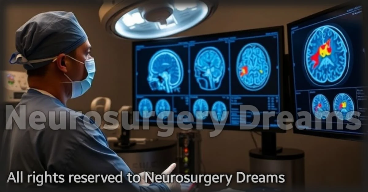
The Future of Brain Mapping: How Advanced Imaging Techniques Are Revolutionizing Neurosurgery
In the rapidly advancing world of neurosurgery, one of the most exciting developments is the evolution of brain mapping techniques. These advanced imaging technologies, such as functional MRI (fMRI) and Diffusion Tensor Imaging (DTI), are providing neurosurgeons with unprecedented insight into the brain's structure and function. This article will explore how these cutting-edge technologies are revolutionizing brain surgery, improving surgical precision, and enhancing patient outcomes.
The Importance of Brain Mapping in Neurosurgery
Brain mapping is the process of creating detailed maps of the brain’s structure and function. This information is essential for neurosurgeons to understand the complex network of brain regions responsible for different functions, such as movement, sensation, and speech. By utilizing advanced imaging techniques, surgeons can pinpoint critical areas in the brain that must be preserved during surgery, reducing the risk of neurological damage.
How Brain Mapping Helps Neurosurgeons
Brain mapping plays a vital role in various aspects of neurosurgery:
- Pre-surgical Planning: Before surgery, detailed brain maps help identify areas that are responsible for vital functions. This allows surgeons to plan the safest surgical approach and avoid damaging crucial brain structures.
- Tumor Localization: Brain mapping enables neurosurgeons to precisely locate tumors, even those hidden deep within the brain, helping them decide the optimal surgical approach for removal.
- Functional Preservation: By understanding the functional organization of the brain, surgeons can avoid disrupting regions that control essential abilities, such as speech, memory, or motor functions.
Advanced Brain Mapping Techniques
In recent years, several groundbreaking brain mapping techniques have emerged, each providing a unique and detailed view of the brain. These technologies offer enhanced accuracy and allow for a more individualized approach to brain surgery.
Functional MRI (fMRI)
Functional MRI is a non-invasive imaging technique that measures brain activity by detecting changes in blood flow. It provides real-time images of brain activity, allowing neurosurgeons to identify which areas of the brain are active during specific tasks, such as speaking or moving. This helps surgeons avoid areas that are crucial for normal function while performing surgery.
Diffusion Tensor Imaging (DTI)
Diffusion Tensor Imaging is an advanced MRI technique that visualizes the brain’s white matter pathways, which are responsible for communication between different brain regions. This allows neurosurgeons to map out critical neural pathways and ensure they are preserved during surgery, particularly when working in areas of the brain involved in movement or cognition.
Magnetoencephalography (MEG)
Magnetoencephalography is a technique that measures the magnetic fields generated by neuronal activity. It is used to map brain function in real-time, helping to pinpoint areas responsible for motor functions, language, and other critical abilities. MEG can be combined with other imaging techniques to provide a comprehensive view of brain activity during surgery.
How These Techniques Improve Patient Outcomes
The integration of advanced brain mapping techniques into neurosurgery has led to significant improvements in patient outcomes:
- Increased Surgical Precision: Surgeons can navigate the brain with greater accuracy, minimizing damage to healthy tissue and reducing the likelihood of postoperative complications.
- Faster Recovery: By preserving essential brain functions and minimizing damage during surgery, patients experience quicker recoveries and fewer neurological deficits.
- Better Quality of Life: With improved surgical outcomes, patients are more likely to regain full function, leading to a higher quality of life after surgery.
The Future of Brain Mapping in Neurosurgery
As technology continues to advance, the future of brain mapping in neurosurgery looks even more promising. The development of new imaging techniques, combined with artificial intelligence (AI), will allow for even more accurate and personalized surgeries. AI-powered systems may analyze brain scans to automatically identify critical areas, further enhancing surgical precision and safety.
Additionally, with the growing potential for augmented reality (AR) and real-time brain mapping during surgery, surgeons will be able to interact with 3D models of the brain while operating, offering unparalleled accuracy and reducing the risk of errors.
Conclusion
Advanced brain mapping techniques are revolutionizing the field of neurosurgery, enabling surgeons to plan and execute procedures with remarkable precision. These technologies are improving patient outcomes, reducing risks, and enhancing the overall surgical experience. As new advancements in imaging and AI continue to emerge, the future of neurosurgery is brighter than ever, offering hope for patients undergoing brain surgery worldwide.

